Abstract
Introduction . Cytogenetic abnormalities (CA) detected by iFISH are among the most powerful prognostic factors in multiple myeloma (MM). With the advent of an unpreceded therapeutic armamentarium during the last years, a thoroughly assessment of individual patients characteristics is necessary in order to choose the best therapy. Among the former, age is an important biologic feature that stratifies patients with regard to treatment intensity.
Aims . To study the distribution of individual CA by iFISH according to different age groups and to evaluate the relationship between age and different cytogenetic profiles.
Materials and methods . We retrospectively evaluated 1116 patients (pts) analyzed in our laboratory between 01/2010 and 12/2016 for whom baseline cytogenetics by iFISH was available. iFISH was carried out on CD138 enriched plasma cells combined with immunofluorescent detection of light chain (LC) restricted plasma cells. The following CA were considered: gain(1q21), gain(9q34), del(13q14), del(17p13), t(4;14), t(11;14), t(14;16). Commercially available DNA probes were used according to manufacturer protocols (MetaSystems, Vysis). In house cutoff levels of 15% and 17% were set for chromosomal imbalances and IgH translocations respectively. Patients were stratified into 3 age groups: 1st group ≤ 45 years (y), 2nd group > 45y and ≤ 65y and 3th group > 65y. High risk cytogenetics (HRC) were defined according to IMWG criteria as at least one of the following by iFISH: del(17p), t(4;14) and t(14;16). Pairwise comparisons between subgroups were performed by the Mann-Whitney or Kruskal Wallis test for continuous variables and by Fisher's exact test for categorical variables. All statistical analyses were conducted in R, version 3.0.1.
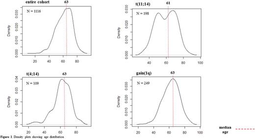
Results . The 1st group comprised 92 (8%) pts, the 2nd 581 (52%) pts and the 3rd 443 (40%) pts. The t(4;14) was detected in 109 (9.8%) pts with higher prevalence in the 2nd and 3rd groups (7.6% vs 10.3% vs 9.5%, p=0.67). The t(11;14) was detected in 198 (18%) pts with a higher prevalence in younger pts (26% vs 18 % vs 16%, p=0.15); age density plots showed two prevalence peaks at 45y and 65y respectively (Fig 1). The t(14;16) was identified in 26 pts (2.3%) and occurred more frequent under 65y (2.7% vs 4.2% vs 1.6%, p=0.11). Gain(9q) was identified in 365 (32%) of 781 evaluable patients occurring less frequently under the age of 45y (22% vs 35% vs 31%, p=0.4). Gain(1q) was detected in 249 (22%) pts with a trend towards higher prevalence in pts over 45y (14% vs 23% vs 23%, p=0.09). Del(17p) was detected in 153 pts (14%) slightly more frequent in the 2nd age group (13% vs 15% vs 12%, p=0.4). Del(13q) was more frequent in the 2nd group (32% vs 42% vs 37%, p=0.12).
Next, we analyzed the distribution of HRC according to age category and identified 36 (21%) pts in the 1st group, 188 (26%) pts in the 2nd group and 119 (22%) pts in the 3rd group (p= 0.21). If gain(1q) was added to the HRC profile, the distribution of the HRC between groups differed significantly (27% vs 40% vs 34%, p=0.012).
For the entire cohort, we further looked how t(4;14) and t(11;14) co-occur with gain(1q) and del(17p). Overall, 38% of the patients with t(4;14) had a gain(1q) (OR 2.8, p< 0.001). Conversely, no significant relation was detected for t(11;14) and for both translocations and del(17p). An inverse relation was found for del(17p) and gain(1q), with only 22% of pts with del(17p) harboring gain(1q) (OR 0.59, p=0.03). An interesting observation was the distribution of the k and λ LC across different CA. We found a significantly higher prevalence of k LC in MM with t(11;14) (60%, p=0.015), gain(1q) (54%, p=0.001) and HRC (67%, p<0.001). Regarding age, no significant difference was identified for the frequency of one type LC or another between age groups (p=0.978).
Conclusion. In line with previous data,our study on a large unselected patient cohort suggests an age dependent distribution of CA. Though not reaching statistical significance, t(11;14) tends to occur more often in younger pts. The HRC profile is found more frequently in pts between 45y and 65y . Adding gain(1q) to the HRC panel delineates this group more clearly. Still, the prognostic value of 1q in the era of novel agents is controversial and might be related to the strong association with t(4;14).
Knop: Takeda: Consultancy.
Author notes
Asterisk with author names denotes non-ASH members.


This feature is available to Subscribers Only
Sign In or Create an Account Close Modal